Scanning Electronic Microscopy
Staff
Scientific Responsible: Dr. Xavier Turon (xturon@ceab.csic.es)
Technical Responsible: Maria García (maria@ceab.csic.es)
Introduction to the service
The electronic microscopy service is available for the observation of samples with the Scanning Electron Microscope (SEM) and for energy dispersive X-ray microanalysis (EDS).
All the necessary equipment for sample preparation is available and the service gives advice to users concerning both sample preparation and the use of the different devices. In addition, forms are available at the service for processing potential suggestions or complaints in order to improve it.


Applications
The use of SEM has a wide range of possibilities:
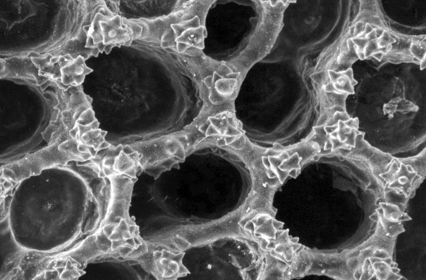
- Structure and ultrastructure of animal and plant tissues and organs.
- Forensic studies (search of particles, tissues, threads, semen…)
- Identification of minerals, synthetic substances, antibiotics, etc.
- Studies in metal and alloys corrosion
- Biodeterioration of works of art.
- Rock and mineral texture
In addition, fundamental microanalysis allows the characterization of the chemical elements present in different parts of a sample.
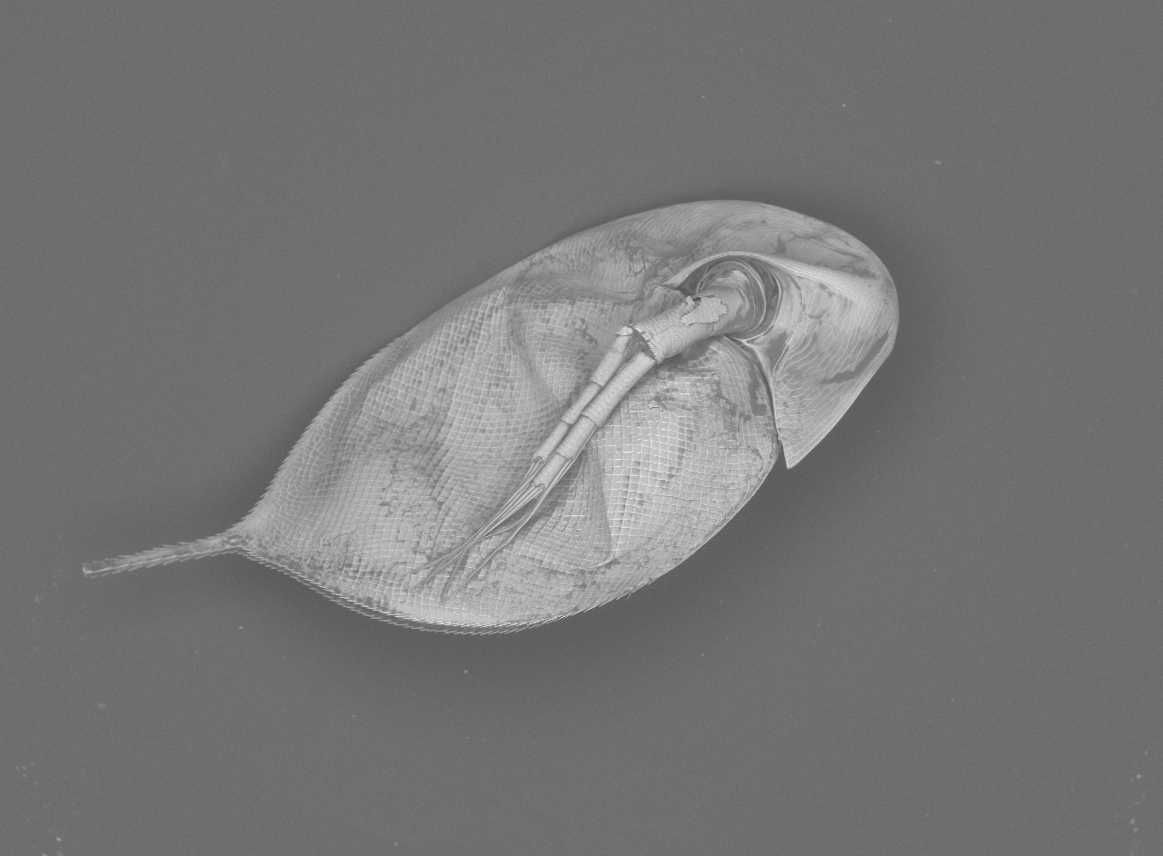
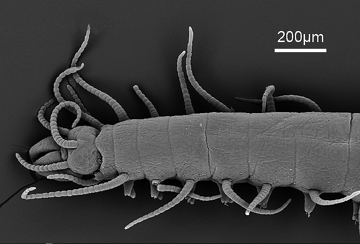
We are currently using this technique mainly in the field of taxonomy with the observation of the relevant specific features of distinct groups of organisms (sponges, polychaetes, macro-invertebrates, etc.) as well as for other studies related to symbiosis and parasitism.
Techniques
Biological samples observed at the SEM service are habitually preserved via freezing or by fixative substances (formaldehyde, alcohol, etc.) and can reach a maximum size of 70 mm in diameter and 50 mm in thickness. Samples can be observed at half vacuum if they have not been dehydrated or in full vacuum if they are completely dehydrated. For dehydration, a treatment is performed at the laboratory by means of hexamethyldisilazane which completely eliminates water.
Devices and characteristics
Scanning Electron Microscope HITACHI-TECH, model TM3000 TABLETOP
Magnification: 15-30.000x (digital zoom 2x, 4x)
Acceleration Tension: 5Kv-15Kv-Analyse
Observation mode: standard or with charge-up reduction
Maximum sample size: 70 mm (diameter)
Maximum sample thickness: 50 mm
Image displacement: +/- 50 um (method 15 kV: D = 4.5 mm)
Frequency: 50/60 Hz
Digital resolution of the image up to 1280 960 pixels
Cooling sample holder HAN (up to -25ºC)
3D-Image viewer
X-ray BRUKER microanalyser
Software: QUANTAX70
Spectrometer EDS
Qualitative and quantitative analysis


Devices for sample preparation
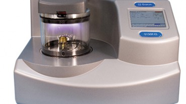
Sputter coater: QUORUM TECHNOLOGIES Q150R/S/E/ES
Information to users
- SAMPLE CHARACTERISTICS AND DELIVERY PROCEDURE
Samples should be sent stating reception method, treatments to be done, name of the user delivering the samples, date, technician receiving the samples and results delivery date.- RESULTS DELIVERY DEADLINE
- Results and rapports will be delivered to the user via e-mail, and security copies will be stored by triplicate (e-mail, hard drive of the Microscope PC and external hard drive). In addition, sample names, characteristics and treatments performed will be stored in the standardised laboratory notebook (CSIC).
- MAXIMUM RESULT STORAGE TIME
- Results will be stored for 5 years.
- SAMPLE MANAGEMENT
- Once the results are delivered and approved by the user, the CEAB will not keep the sample surpluses unless the user asks for it.
- COMMUNICATION SERVICE-USER
- It will take place via e-mail and phone. Persons to contact with are the scientific and the technical responsible of the Service.
- Service satisfaction survey
complete the form